We spoke. Now the next topic is the shoulder muscles. To pump up powerful shoulders, you need to know how to pump up, how to distribute efforts and create a harmonious shape. Not the least important thing is anatomy.
Knowing how our muscles work means achieving our plans in less time and without physical losses. And targeted exercises will more accurately lead to the planned results.
Swing, shoulder, spread, arm...
Our shoulder is designed in such a way that two muscle groups are considered - the anterior and posterior groups. They perform flexion and extension functions, respectively.
They cover tendons, bones, blood vessels, and connect the arms to the body. They protect the shoulder from injury, help the arms move in different directions, and the elbow bend.
The anterior flexion group consists of:
- coracobrachial;
- biceps brachii;
- brachial muscle.
The posterior extensor muscle is represented by:
- triceps brachii;
- elbow muscle.
Before planning training, I recommend that novice bodybuilders familiarize themselves with the Human Atlas and study in detail the anatomical structure of the attachment of the muscles of the forearm and shoulder girdle.
You will see the muscles of the chest, upper back, neck, and muscles that act on the elbow joint in this section. Each muscle has a name and is endowed with a specific responsibility for the freedom of movement of the limb.

For example, we are talking about things like:
- deltoid;
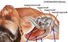
- supraspinatus;
- infraspinatus;
- round small and large muscles;
- subscapular.
How to train your shoulder correctly
Beautiful deltoids are the pride of an athlete. They attract the attention and admiration of others. The results of hard training, sometimes with injuries, when you have to overcome pain, long months of recovery and continued work on yourself are obvious.
The shoulder joint reacts sharply to uncalculated physical activity or incorrect exercises and is difficult to treat.
Please note that the shoulder muscles are involved in all basic exercises with and without loads, no matter what other muscles the athletes exercise. In this case, the pressing functions are performed by the anterior muscle bundles, and the traction functions are performed by the rear deltoids.

Of all the exercises, the most effective in imparting strength to the shoulder muscles is the standing barbell press, and for volume and mass, the barbell row to the chin.
To work out the muscle tissue of the shoulders, take one or two times a week. Do not exercise on cold muscles. A mandatory warm-up will save your health, time to prepare for competitions, and save muscles and tendons from damage.
In the first half of the training, basic exercises are usually performed, in the second half they work on the shoulder itself. Here you need to focus on vertical presses and isolation exercises (two or three will be enough), such as pull-downs and lateral raises.

Perform vertical presses with a barbell or dumbbells in three to four sets of 6-12 repetitions. While isolating - in two or three sets of 10-15 repetitions. Start with low loads and increase them as you gain experience and strengthen your shoulder muscles.
Shoulder injuries
Take your time to get results here and now. Let it be a long road, but stable. Shoulder injuries can occur due to displacement of the head of the humerus during a sudden jerk of the barbell. Do not test the tendons for rupture with heavy weights.
Often muscles hurt from overload. Give them a rest. After all, during rest, muscle mass grows.
Almost all basic exercises for strength and volumetric muscle strengthening are dangerous. You should remember this and develop rules for yourself:
- bench press;
- overhead barbell press;
- dumbbell raises, tilted to the sides;
- raising dumbbells to the sides while lying on your back;
- cravings for the chest.

First, take care to choose the right weight. It is because of excessive loads that tendons tear, sprained ligaments cause pain and the inability to continue training. Shoulder dislocation is one of the most common bodybuilder injuries when the head of the humerus protrudes anteriorly.
Dislocation is accompanied by sharp pain and crunching. It is advisable not to repair the injury yourself; let a professional doctor do it. If you experience any discomfort or pain, do not continue training by force.
Recovery takes 10 to 14 days, during which time the joint must be left alone. You can resume training after the pain disappears. This should be done sparingly in relation to the muscles.
At first these will be warm-up movements without weights. Then, over the course of a month and a half, gradually increase the load, based on the sensations.

Remember these three main rules, the implementation of which will bring you benefit and satisfaction from hard work: basic multi-joint exercises, high-calorie nutrition, weekly analysis of what you have done.
Broad shoulders and health to everyone! Subscribe to updates on my blog, share with friends and friends of friends on social networks. Until the next topics on my page.
Front view.
deltoid muscle (turned posteriorly);
pectoralis minor muscle (cut off);
levator scapulae muscle (severed);
subscapularis muscle;
three-way hole;
teres major;
latissimus dorsi (cut off);
coracobrachialis muscle;
long head of the triceps brachii muscle;
medial head of the triceps brachii;
brachialis muscle;
medial epicondyle of the humerus;
aponeurosis of the biceps brachii muscle;
fascia of the forearm;
brachioradialis muscle;
biceps brachii tendon;
pronator teres;
biceps brachii;
short head of the biceps brachii muscle;
large pectoral muscle;
tendon of the long head of the biceps brachii muscle
The deltoid muscle (m. deltoideus) (Fig. 90, 101, 104, 106, 111, 112, 113, 114) moves the shoulder outward to a horizontal plane, while the front bundles of the muscle pull the arm forward, and the rear bundles back. It is a thick, triangular-shaped muscle that covers the shoulder joint and parts of the shoulder muscles. Its large bunches fan-shapedly converge to the apex of the triangle, directed downward. The muscle starts from the axis of the scapula, acromion and lateral part of the clavicle, and is attached to the deltoid tuberosity of the humerus. Under the lower surface of the muscle is the subdeltoid bursa (bursa subdeltoidea).
The supraspinatus muscle (m. supraspinatus) (Fig. 102, 114) has a triangular shape and lies in the supraspinatus fossa of the scapula, located directly under the trapezius muscle. The supraspinatus muscle lifts the shoulder and retracts the capsule of the shoulder joint, preventing it from pinching. The origin of the muscle is on the surface of the supraspinatus fossa, and the attachment point is on the upper platform of the greater tubercle of the humerus and on the posterior surface of the capsule of the shoulder joint.
The infraspinatus muscle (m. infraspinatus) (Fig. 101, 102, 104, 114) turns the shoulder outward, pulls the raised arm back and pulls the capsule of the shoulder joint. This is a flat, triangular-shaped muscle that fills the entire infraspinatus fossa. Its upper part is covered by the trapezius and deltoid muscles, and the lower part by the latissimus dorsi and teres major muscles. The infraspinatus muscle starts from the wall of the infraspinatus fossa and the posterior surface of the scapula, and is attached to the middle platform of the greater tubercle of the humerus and the capsule of the shoulder joint. At the point of its attachment to the humerus there is a subtendinous bursa of the infraspinatus muscle (bursa subtendinea mm. infraspinati).
a) long head, b) medial head;
12 - biceps brachii;
13 - brachial muscle;
14 - pronator teres;
15 - aponeurosis of the biceps brachii muscle;
16 - brachioradialis muscle;
17 - fascia of the forearm

a) short head, b) long head;
2 - deltoid muscle;
3 - subscapularis muscle;
4 - coracobrachialis muscle;
5 - teres major muscle;
6 - triceps brachii muscle: a) long head, b) medial head;
7 - brachialis muscle;
8 - biceps brachii tendon

side view
1 - supraspinatus fascia;
2 - infraspinatus fascia;
3 - teres major muscle;
4 - deltoid muscle;
5 - triceps brachii muscle: a) long head, b) lateral head, c) medial head;
6 - biceps brachii;
7 - brachialis muscle;
8 - triceps brachii tendon;
9 - brachioradialis muscle;
10 - extensor carpi radialis longus;
12 - fascia of the forearm

back view
1 - supraspinatus fascia;
2 - supraspinatus muscle;
3 - infraspinatus fascia;
4 - infraspinatus muscle;
5 - teres minor muscle;
6 - teres major muscle;
7 - deltoid muscle;
8 - triceps brachii muscle: a) long head, b) lateral head, c) medial head;
9 - triceps brachii tendon;
10 - brachioradialis muscle;
11 - extensor carpi radialis longus;
13 - fascia of the forearm
The teres minor muscle (m. teres minor) (Fig. 101, 102, 104, 114) turns the shoulder outward, at the same time slightly moving it back, and retracts the capsule of the shoulder joint. An oblong, rounded muscle, the upper part of which is adjacent to the infraspinatus muscle, the anterior part is covered by the deltoid muscle, and the posterior part is covered by the teres major muscle. The point of origin is located on the posterior surface of the scapula below the infraspinatus muscle, and the attachment point is on the lower platform of the greater tuberosity of the humerus and the posterior surface of the capsule of the shoulder joint.
0The large round muscle (m. teres major) (Fig. 101, 104, 105, 112, 113, 114) turns the shoulder inward and pulls it back, bringing the arm to the body. An oblong flat muscle adjacent to the latissimus dorsi muscle and partially covered by it in the posterior section. In the outer section, the teres major muscle is covered by the deltoid muscle. The starting point is the posterior surface of the scapula at its lower angle, the attachment point is the crest of the lesser tubercle of the humerus. Near the attachment site is the subtendinous bursa of the teres major muscle (bursa subtendinea mm. teretis majoris).
The subscapularis muscle (m. subscapularis) (Fig. 105, 111, 112) rotates the shoulder inward and takes part in its adduction to the body. A flat, triangular-shaped vastus muscle that fills the entire subscapularis fossa. It begins on the surface of the subscapularis fossa and ends on the lesser tubercle of the humerus and on the anterior surface of the capsule of the shoulder joint.
At the attachment site there is a small subtendinous bursa of the subscapularis muscle (bursa subtendinea mm. subscapularis)
The shoulder muscles are divided into anterior (mainly flexors) and posterior (extensor) groups.
Front group
The biceps brachii muscle (m. biceps brachii) (Fig. 90, 106, 111, 112, 113, 115, 116, 117, 124) flexes the forearm at the elbow joint and rotates it outward, raising the arm. A rounded fusiform muscle consisting of two heads (due to the long head (caput longum) the arm is abducted, thanks to the short head (caput breve) it is adducted) and is located in the area of the shoulder and elbow bend directly under the skin. The long head starts from the supraglenoid tubercle of the scapula, and the short head starts from the coracoid process of the scapula.
The heads unite and form a common abdomen, which is attached to the tuberosity of the radius. Part of the fibrous bundles is directed medially, forms a lamellar process, which is called the aponeurosis of the biceps brachii muscle (aponeurosis m. bicipitis brachii) (Fig. 111, 115) and passes into the fascia of the forearm.
The coracobrachialis muscle (m. coracobrachialis) (Fig. 111, 112) raises the shoulder and brings the arm to the midline. A flat muscle covering the short head of the biceps brachii muscle. Its point of origin is at the apex of the coracoid process of the scapula, and its attachment point is just below the middle of the medial surface of the humerus. Near the point of origin is the coracohumeral bursa (bursa mm. coracobrachialis).
The brachialis muscle (m. brachialis) (Fig. 90, 111, 112, 113, 115, 116, 124) flexes the shoulder and tightens the capsule of the shoulder joint. The muscle is wide, fusiform, located on the anterior surface of the lower half of the shoulder under the biceps muscle. It begins on the outer and anterior surface of the humerus and is attached to the tuberosity of the humerus, as well as partially to the capsule of the elbow joint.
Back group
The triceps brachii muscle (m. triceps brachii) (Fig. 90, 101, 104, 111, 112, 113, 114, 118, 124) extends the forearm, thanks to its long head, pulls the arm back and brings the shoulder to the body. A long muscle located on the entire back surface of the shoulder from the scapula to the olecranon. The long head (caput longum) begins on the subarticular tubercle of the scapula, the lateral head (caput laterale) - on the posterolateral surface of the humerus from the greater tubercle above the radial groove, the medial head (caput mediale) - on the posterior surface of the humerus below the radial groove, it is partially covered long and lateral heads. All three heads form a fusiform belly, which passes into the tendon and attaches to the olecranon process and the capsule of the elbow joint.
The elbow muscle (m. anconeus) (Fig. 90, 113, 114, 118) extends the forearm at the elbow joint, retracting the capsule of the elbow joint. The muscle is a continuation of the medial head of the triceps brachii muscle and has a pyramidal shape. Its point of origin is located on the lateral epicondyle of the humerus, and its attachment point is on the olecranon process and the posterior surface of the body of the ulna.
Muscles of the anterior abdominal wall
The rectus abdominis (m. rectus abdominis) (Fig. 90, 109, 110) tilts the torso anteriorly. It is part of the abdominal press and provides intra-abdominal pressure, due to which the internal organs are held in a certain position. In addition, she takes part in the acts of urination, defecation and childbirth. This long, flat muscle is located in the anterior abdominal wall on the sides of the white line (linea alba), which runs from the xiphoid process of the sternum to the pubic fusion. The point of origin of the rectus abdominis muscle is located on the xiphoid process of the sternum and the cartilages of the V-VII ribs, and the attachment point is on the pubic bone between the pubic tubercle and the pubic symphysis (symphysis). The muscle bundles of the rectus abdominis muscle are interrupted by three to four transversely located tendon bridges, two of which are located above the navel, the third at the level of the navel, and the fourth (poorly developed) below.

Muscles of the anterior wall of the abdomen and pelvis
1 - rectus abdominis muscle;
2 - fascia iliaca;
3 - iliopsoas muscle;
4 - interfoveal ligament;
5 - external iliac artery;
6 - external iliac vein;
7 - internal locking muscle;
8 - muscle that lifts the ani;
9 - external locking muscle
The pyramidal muscle of the abdomen (m. pyramidalis) (Fig. 90, 110) stretches the linea alba. The muscle has a triangular shape, begins on the pubic bone, anterior to the insertion of the rectus abdominis muscle, and is attached at various levels of the lower part of the linea alba.
Muscles of the anterior surface of the human body

General form.
1 - trapezius muscle;
sternocleidomastoid muscle;
depressor anguli oris muscle;
masticatory muscle;
zygomaticus major;
orbicularis oculi muscle;
temporal muscle;
anterior belly of the supracranial muscle,
orbicularis oris muscle;
muscle that depresses the lower lip;
deltoid,
biceps brachii;
rectus abdominis muscle;
external oblique abdominal muscle;
pyramidal muscle;
pectineus muscle;
long adductor muscle of the thigh;
sartorius;
adductor magnus muscle of the thigh;
rectus femoris muscle;
vastus medialis;
tibialis anterior;
tendons of the long extensor toe muscle;
soleus muscle;
calf muscle;
vastus lateralis femoris;
tensor fascia lata muscle;
muscle that extends the fingers;
long radialis muscle, extensor carpi;
brachioradialis muscle;
brachialis muscle;
serratus anterior;
large food muscle.
Muscles of the hand, right. Palmar side

muscle - pronator quadratus;
flexor carpi ulnaris tendon;
pisiform bone;
flexor tendon retinaculum;
muscle opposite the little finger;
6 and palmar interosseous muscles;
vermiform muscle (cut off);
deep transverse metacarpal ligament;
flexor digitorum superficialis tendon (severed);
deep digital flexor tendon;
fibrous tendon sheath;
I dorsal interosseous muscle;
adductor pollicis muscle;
tendon of the flexor pollicis longus muscle;
muscle - short flexor of the thumb;
muscle that opposes the thumb;
tendon of the abductor pollicis longus muscle;
muscle, flexor pollicis longus.
Muscles of the hand

The muscles of the hand are located mainly on the palmar surface of the hand and are divided into the lateral group (muscles of the thumb), medial group (muscles of the little finger) and the middle group. On the dorsum of the hand are the dorsal (back) interosseous muscles.
Lateral group
The short muscle that abducts the thumb (m. abductor pollicis brevis) (Fig. 120, 121) abducts the thumb, slightly opposing it, and takes part in flexion of the proximal phalanx. It is located directly under the skin on the lateral side of the eminence of the thumb. It begins on the scaphoid bone and ligament of the palmar surface of the wrist, and is attached to the lateral surface of the base of the proximal phalanx of the thumb.
a) belly, b) tendon;
3 - muscle opposing the thumb;
4 - flexor retinaculum;
5 - flexor pollicis brevis;
6 - short muscle, abductor pollicis;
7 - muscle adducting the little finger;
8 - palmar interosseous muscles;
9 - adductor pollicis muscle: a) oblique head, b) transverse head;
10 - vermiform muscle;
11 - dorsal interosseous muscle;
12 - superficial digital flexor tendon;
13 - sheath of the tendons of the fingers;
14 - deep digital flexor tendon
palmar surface
1 - pronator quadratus;
2 - tendon of the brachioradialis muscle;
3 - flexor carpi ulnaris tendon;
4 - flexor carpi radialis tendon;
5 - muscle opposing the thumb;
6 - flexor pollicis brevis;
7 - palmar interosseous muscles;
8 - short muscle, abductor pollicis;
9 - dorsal interosseous muscles

The short flexor of the thumb (m. flexor pollicis brevis) (Fig. 116, 120, 121) flexes the proximal phalanx of the thumb. This muscle is also located just under the skin and has two heads. The starting point of the superficial head is on the ligamentous apparatus of the palmar surface of the wrist, and the deep head is on the trapezius bone and the radiate ligament of the wrist. Both heads are attached to the sesamoid bones of the metacarpophalangeal joint of the thumb.
The muscle opposing the thumb to the hand (m. opponens pollicis) (Fig. 116, 120, 121) opposes the thumb to the little finger. It is located under the abductor pollicis brevis muscle and is a thin triangular plate. The muscle starts from the ligamentous apparatus of the palmar surface of the wrist and the tubercle of the costoptrapezium, and is attached to the lateral edge of the first metacarpal bone.
The muscle that adducts the thumb (m. adductor pollicis) (Fig. 120, 123) adducts the thumb and takes part in the flexion of its proximal phalanx. It lies the deepest of all the muscles of the eminence of the thumb and has two heads. The starting point of the transverse head (caput transversum) is located on the palmar surface of the IV metacarpal bone, the oblique head (caput obliquum) is on the capitate bone and the radiate ligament of the wrist. The attachment point for both heads is located at the base of the proximal phalanx of the thumb and the medial sesamoid bone of the metacarpophalangeal joint.
Medial group
The short palmaris muscle (m. palmaris brevis) (Fig. 115) stretches the palmar aponeurosis, forming folds and dimples in the skin in the area of the eminence of the little finger. This muscle, which is a thin plate with parallel fibers, is one of the few cutaneous muscles available in humans. It has a point of origin on the inner edge of the palmar aponeurosis and the ligamentous apparatus of the wrist. The place of its attachment is located directly in the skin of the medial edge of the hand at the eminence of the little finger.
The muscle that abducts the little finger (m. abductor digiti minimi) (Fig. 122, 123) abducts the little finger and takes part in the flexion of its proximal phalanx. It is located under the skin and is partially covered by the palmaris brevis muscle. The muscle originates from the pisiform bone of the wrist and attaches to the ulnar edge of the base of the proximal phalanx of the little finger.
The short flexor of the little finger (m. flexor digiri minimi) bends the proximal phalanx of the little finger and takes part in its adduction. It is a small, flattened muscle covered by skin and partly by the palmaris brevis muscle. Its point of origin is located on the hamate and ligaments of the wrist, and its attachment point is on the palmar surface of the base of the proximal phalanx of the little finger.
The muscle that adducts the little finger (m. opponens digiti minimi) (Fig. 116, 120) opposes the little finger to the thumb. The outer edge of the muscle is covered by the short flexor of the little finger. It begins on the hamate and ligamentous apparatus of the wrist, and is attached to the ulnar edge of the fifth metacarpal bone.

back surface
1 - short extensor pollicis;
2 - extensor of the little finger;
3 - extensor carpi ulnaris tendon;
4 - extensor finger;
5 - extensor carpi radialis longus tendon;
6 - tendon of the short extensor carpi radialis;
7 - tendon of the long extensor pollicis;
8 - extensor tendon of the little finger;
9 - muscle that abducts the little finger;
10 - extensor tendon;
11 - extensor tendon of the index finger;
12 - dorsal interosseous muscles;
13 - flexor pollicis longus tendon
The muscles of the free part of the lower limb are divided into thigh muscles, leg muscles and foot muscles.
The thigh muscles surround the thigh bone and are divided into an anterior muscle group, which consists primarily of extensors, a medial group, which includes adductors, and a posterior muscle group, which includes flexors.
Front group
The sartorius muscle (m. sartorius) (Fig. 90, 129, 132, 133, 134, 145) flexes the thigh and lower leg, simultaneously rotating the thigh outward and the lower leg inward, providing the ability to cross legs. It is a narrow ribbon, located on the front surface of the thigh and, spiraling down, passes to the front surface. The sartorius muscle is one of the longest muscles in humans. It starts from the superior anterior iliac spine, and is attached to the tibial tuberosity and in separate bundles to the fascia of the leg.
Image

Rice. 131. Muscles of the pelvis and thigh (front view):
1 - piriformis muscle;
2 - gluteus minimus;
3 - external locking muscle;
4 - quadriceps femoris muscle;
5 - short adductor muscle;
6 - adductor magnus;
7 - vastus lateralis muscle;
8 - adductor channel
Image

Rice. 132. Muscles of the pelvis and thigh (side view):
1 - psoas major muscle;
2 - iliacus muscle;
3 - piriformis muscle;
4 - internal locking muscle;
5 - pectineus muscle;
6 - gluteus maximus muscle;
7 - long adductor muscle;
8 - adductor magnus;
9 - sartorius muscle;
10 - thin muscle;
11 - semitendinosus muscle;
13 - semimembranosus muscle;
14 - vastus medialis;
15 - calf muscle
The quadriceps femoris (m. quadriceps femoris) (Fig. 131) consists of four heads and is the largest human muscle. When all heads contract, it extends the lower leg; when the rectus femoris contracts, it takes part in its flexion. It is located on the anterolateral surface of the thigh, in the lower parts it completely passes to the lateral surface. Each of the heads has its own starting point. The longest rectus femoris muscle (m. rectus femoris) (Fig. 90, 129, 132, 145) begins on the lower anterior iliac spine; vastus medialis (m. vastus medialis) (Fig. 90, 129, 130, 132, 133, 145) - on the medial lip of the linea aspera of the femur; vastus lateralis (m. vastus lateralis) (Fig. 90, 129, 130, 131, 133, 145) - on the greater trochanter, intertrochanteric line and lateral lip of the linea aspera of the femur; intermediate broad muscle of the thigh (m. vastus intermedius) (Fig. 130, 145) - on the anterior surface of the femur. All heads grow together to form a common tendon, which is attached to the apex and lateral edges of the patella, past which the tendon descends below and passes into the patellar ligament, which is attached to the tibial tuberosity. At the site of muscle attachment there is the bursa suprapatellaris, subcutaneous prepatellaris, bursa subcutanea infrapatellaris, and deep subpatellar bursa.
The articular muscle of the knee (m. articularis genus) (Fig. 136) stretches the bursa of the knee joint. It is a flat plate and is located on the front surface of the thigh under the vastus intermedius muscle. Its starting point is on the anterior surface of the lower third of the femur, and its attachment point is on the anterior and lateral surfaces of the articular capsule of the knee joint.
Medial group

The pectineus muscle (m. pectineus) (Fig. 90, 129, 130, 132) flexes and adducts the thigh, rotating it outward. The flat muscle is quadrangular in shape, originates on the crest and superior ramus of the pubis, and is inserted on the medial lip of the linea aspera of the femur below the lesser trochanter.
The thin muscle (m. gracilis) (Fig. 90, 129, 130, 132, 134, 145) adducts the thigh and takes part in flexing the tibia, turning the leg inward. The long, flat muscle is located just under the skin. Its point of origin is on the lower branch of the pubic bone, and its attachment point is on the tuberosity of the tibia. The gracilis tendon fuses with the sartorius and semitendinosus tendons and the fascia of the leg to form the superficial pes anserine. The so-called goose bursa (bursa anserina) is also located here.
Image
Rice. 133. Muscles of the pelvis and thigh (side view):
1 - latissimus dorsi muscle;
2 - external oblique abdominal muscle;
3 - gluteus medius muscle;
4 - gluteus maximus muscle;
5 - sartorius muscle;
6 - muscle that tightens the fascia lata of the thigh;
7 - iliotibial tract;
9 - biceps femoris muscle: a) long head, b) short head;
10 - vastus lateralis muscle;
11 - calf muscle
Image

Rice. 134. Muscles of the pelvis and thigh (rear view):
1 - gluteus maximus muscle;
2 - adductor magnus;
3 - iliotibial tract;
4 - tendon jumper of the semitendinosus muscle;
5 - semitendinosus muscle;
6 - biceps femoris muscle;
7 - thin muscle;
8 - semimembranosus muscle;
9 - sartorius muscle;
10 - plantaris muscle;
11 - gastrocnemius muscle: a) medial head, b) lateral head
The long adductor muscle (m. adductor longus) (Fig. 90, 129, 130, 132) adducts the thigh and takes part in its flexion and outward rotation. This is a flat muscle, shaped like an irregular triangle and located on the anteromedial surface of the thigh. It starts from the superior ramus of the pubis and inserts on the middle third of the medial lip of the linea aspera of the femur.
The short adductor muscle (m. adductor brevis) (Fig. 131) adducts the thigh and takes part in its flexion and outward rotation. It is a triangular-shaped muscle that originates on the anterior surface of the inferior ramus of the pubis, lateral to the gracilis muscle, and is inserted on the upper third of the medial lip of the linea aspera of the femur.
The large adductor muscle (m. adductor magnus) (Fig. 129, 130, 131, 132, 134) adducts the thigh, partly rotating it outward. Thick, wide, the most powerful muscle of this group, located deeper than the other adductor muscles. Its point of origin is located on the ischial tuberosity, as well as on the branch of the ischium and the lower branch of the pubic bone. The attachment site is located on the medial lip of the linea aspera and the medial epicondyle of the femur. Several holes are formed in the muscle bundles, allowing blood vessels to pass through. The largest of them is called the tendon opening (hiatus tendineus). Above it is a fascial plate, and between it and the muscle a triangular-shaped space is formed, called the adductor canal (canalis adductorius) (Fig. 131). The femoral vein, artery and hidden nerve of the lower limb pass through it.
Back group
The biceps femoris muscle (m. biceps femoris) (Fig. 133, 134, 145) extends the thigh and flexes the lower leg. In a bent position, rotates the lower leg outward. It runs along the lateral edge of the upper thigh. The muscle has one belly and two heads. The long head (caput longum) starts from the ischial tuberosity, the short head (caput breve) - on the lower part of the lateral lip of the linea aspera of the femur. The abdomen ends in a long narrow tendon, the attachment point of which is on the head of the fibula. Some of the bundles are woven into the fascia of the leg. Near the point of origin of the long muscle is the superior bursa of the biceps femoris muscle (bursa m. bicipitis femoris superior). In the area of the tendon there is the lower subtendinous bursa of the biceps femoris muscle (bursa subtendinea m. bicipitis femoris inferior).
The semitendinosus muscle (m. semitendinosus) (Fig. 130, 132, 134, 145) extends the thigh, bends the lower leg, rotating it inward in a bent position, and also takes part in straightening the torso. The muscle is long and thin, partially covered by the gluteus maximus muscle, sometimes interrupted by a tendon bridge (intersectio tendinea) (Fig. 134). Its point of origin is located on the ischial tuberosity, and its attachment point is on the medial surface of the tibial tuberosity. Individual bundles of muscles are woven into the fascia of the leg, taking part in the formation of the crow's foot.
The semimembranosus muscle (m. semimembranosus) (Fig. 130, 132, 134, 145) extends the thigh and bends the tibia, rotating it inward. It runs along the medial edge of the posterior surface of the thigh and is partially covered by the semitendinosus muscle. The muscle starts from the ischial tuberosity and attaches to the edge of the medial condyle of the tibia.
The tendon is divided into three bundles, forming a deep pes anserine. The external bundle passes into the popliteal fascia, into the posterior ligament of the knee joint.
At the site where the tendon divides into separate bundles there is a synovial bursa of the semimembranosus muscle (bursa m. semimembrano
Shoulder abductor muscles, located on the superolateral surface of the joint: the deltoid surrounds the joint in front, on the lateral side and behind, the supraspinatus lies in the supraspinatus fossa under the trapezius and partly the deltoid muscles. These muscles abduct the shoulder only to a horizontal level.
Shoulder adduction is carried out according to the rule of parallelogram of forces of the muscles located in front and behind the shoulder joint, with their simultaneous contraction: pectoralis major, coracobrachialis (located on the anteromedial surface of the shoulder in its upper section), latissimus dorsi, infraspinatus (lies below the spine of the scapula and partially covered by the deltoid and trapezius muscles), teres minor (adjacent to the lower edge of the infraspinatus muscle), teres major (lies between the teres minor muscle and the upper edge of the latissimus dorsi muscle), subscapularis (located in the subscapularis fossa, it can only be seen on anatomical preparations or in the pictures), the long head of the triceps brachii muscle (located on the back of the shoulder).
Shoulder flexor muscles lie in front of the shoulder joint: the anterior part of the deltoid muscle, pectoralis major, coracobrachialis; The biceps brachii muscle lies under the skin on the front surface of the shoulder.
Shoulder extensor muscles located behind the shoulder joint: the back of the deltoid muscle, infraspinatus, teres minor, teres major, latissimus dorsi, long head of the triceps brachii muscle.
The supinator muscles are attached to the humerus somewhat posteriorly and externally (posterior deltoid, infraspinatus, teres minor), and the pronator muscles are attached to the front (pectoralis major, anterior deltoid, coracobrachialis, subscapularis, teres major, latissimus dorsi) .
If the deltoid and supraspinatus muscles abduct the shoulder to approximately a horizontal level, the coracobrachialis with the long head of the biceps brachii muscle pull the shoulder forward also to the horizontal, then how does the arm rise up to a vertical position? It turns out that none of the muscles of the upper limb girdle and shoulder can produce this movement. To raise the arm to a vertical position, muscle action is required not on the shoulder, but on the scapula, causing it to move around the sagittal axis. The movement is quite complex. First, you need to raise the scapula, which is produced by the levator scapulae muscle. This creates better conditions for the action of the trapezius muscle and the lower teeth of the serratus anterior muscle. When they work together, the lower angle of the scapula shifts to the lateral side and forward, and the lateral angle of the scapula with the glenoid cavity moves upward, which makes it possible for the free upper limb to assume a vertical position.
To make sure of this, you need to feel the lower corner of the scapula in the usual, natural position of the body and mark this point with a dermographic pencil; then raise your hands up and mark the place where the lower angle of the scapula is displaced. It turns out that it shifted laterally and somewhat forward, which led to a displacement of the lateral angle with the glenoid cavity, and then the entire limb upward. This is especially noticeable in thin people.
When studying the biceps brachii muscle, it is necessary to pay attention to the specific course of its tendon through the cavity of the shoulder joint, which determines its effect on this joint and its role in strengthening it. The location of the biceps brachii muscle in relation to the shoulder and elbow joints determines its function: flexion at these joints. But it turns out that the biceps brachii muscle, due to its specific attachment to the tuberosity of the radial bone, is also a strong supinator of the forearm. For comparison, we should recall the single-joint coracobrachialis and brachialis muscles: one acts on the shoulder, the second on the elbow joint.
It is recommended to disassemble in the same way. Of these muscles, the triceps brachii, latissimus dorsi and teres major muscles are of significant importance, especially the second one, when, when lowering the upper limb, it moves it backwards with some pronation of the shoulder (movements of a skier, chopping movements of a fencer, etc.).
When resting on the humerus (the shoulder joint and the girdle of the upper limb are fixed), under the action of the latissimus dorsi and pectoral muscles, the torso is pulled up, for example, when climbing a rope, a pole, or resting on parallel bars.
Holding conferences in Russia: finance, law and taxation, transport. Also on www.ros.biz: analytical articles, news, calendar of events.
1. Deltoid muscle (m. deltoideus)- located directly under the skin, covers the shoulder joint, forms the characteristic roundness of the shoulder.
Starts from the lateral third of the clavicle, acromion, spine of the scapula.
Distinguish three parts: clavicular, acromial and scapular.
The bundles of all parts converge on the outer surface of the humerus and are attached to the deltoid tuberosity of the humerus. Under the deltoid muscle, between the deep plate of its fascia and the greater tubercle of the humerus, there is synovial subdeltoid bursa (bursasubdeltoidea).
Functions:
The clavicular part bends the shoulder, pronates it, and also lowers the raised arm down;
The scapular part extends the shoulder, supinates it, and lowers the raised arm;
The acromion abducts the arm.
2.Supraspinatus muscle (m. supraspinatus)- located in the supraspinous fossa.
It starts from the posterior surface of the scapula above the scapular spine and from the supraspinatus fascia.
Attached to the greater tubercle of the humerus, some of the bundles are woven into the capsule of the shoulder joint.
Functions:
Abducts the shoulder;
3. Infraspinatus muscle (m. Infraspinatus)
It starts from the posterior surface of the scapula below the scapular spine and from the infraspinatus fascia.
Attaches to the greater tubercle of the humerus.
Under the tendon of the infraspinatus muscle is located subtendinous bursa of the infraspinatus muscle (bursa subtendinea musculi infraspinati).
Functions:
Rotates the shoulder outward;
Pulls back the joint capsule, preventing pinching.
4. Teres minor muscle (m. Teres minor)
From the lateral edge of the scapula and infraspinatus fascia to the greater tubercle of the humerus.
Functions:
Rotates the shoulder outward (supination);
Pulls back the joint capsule, preventing pinching.
5. Teres major muscle (m. Teres major)
From the lower part of the lateral border, the lower angle of the scapula and from the infraspinatus fascia to the crest of the lesser tubercle of the humerus.
The tendon of the teres major muscle is adjacent to the tendon of the latissimus dorsi muscle, and is often located between them subtendinous bursa of the latissimus dorsi muscle (bursa subtendinea musculi latissimi dorsi), the lower edges of the tendons of these two muscles are connected over a short distance. Between the tendon of the teres major muscle and the humerus is located subtendinous bursa of the teres major muscle (bursa subtendinea muscul teretis majoris).
Function:
With a fixed scapula, it extends the shoulder in the shoulder joint and simultaneously pronates it;
The raised arm leads to the body;
With a strengthened hand, it pulls the lower corner of the scapula outward and moves it forward.
6. Subscapularis muscle (m. Subscapularis)
From the subscapular fossa and lateral edge of the scapula to the lesser tubercle and crest of the lesser tubercle of the humerus.
Under the tendon of the subscapularis muscle there is subtendinous bursa of the subscapularis muscle.
Functions: turns the shoulder inward at the same time bringing the shoulder towards the body.
Mm. supraspinatus, infraspinatus, subscapularis, teres minor form around the shoulder joint « rotatorycuff", which stabilizes the head of the humerus in the glenoid fossa of the scapula during movements in the shoulder joint. By attaching to the capsule of the shoulder joint, these muscles tighten it and prevent it from being pinched during movement.
The muscles of the upper and lower limbs are divided into groups based on regional affiliation (topography) and the function they perform. Muscles of the upper limb(Fig. No. 87, 88, 89, 90, 91, 92) are usually divided into the muscles of the shoulder girdle and the muscles of the free upper limb: shoulder, forearm and hand. Muscles of the lower limb(Fig. No. 85, 86, 93, 94, 95, 96, 97, 98, 99, 100, 101,102,103) - on the muscles of the pelvic girdle (pelvis) and the free lower limb: thighs, legs and feet. At the same time, a complete analogy cannot be drawn between the muscles of the upper and lower limbs due to the differences in the structure and functions of the girdles and free parts of the limbs. Due to the specific function, the bones of the shoulder girdle are movably connected to the skeleton of the body and have special muscles that act on the collarbone and especially on the scapula. Thanks to this, the scapula and collarbone have great freedom of movement. On the lower limb, the pelvic girdle is firmly, almost motionlessly, connected to the spine at the sacroiliac joint.
For better assimilation of a wide variety of muscles of the limbs, let's consider their graphological structure according to topography and function (see Diagram 1).
Muscles of the shoulder girdle(Fig. No. 83, 87, 88) are located around the shoulder joint and provide it with a full range of movements (with the participation of some muscles of the chest and back). All 6 muscles of this group begin on the bones of the shoulder girdle and attach to the humerus.
1) The deltoid muscle starts from the lateral third of the clavicle, acromion and spine of the scapula. Attaches to the deltoid tuberosity of the humerus. The front part of the muscle flexes the shoulder, the middle part abducts it, and the back part extends the shoulder.
2) The supraspinatus muscle starts from the same-named fossa of the scapula and is attached to the greater tubercle of the humerus. Abducts the shoulder, being a synergist of the middle bundles of the deltoid muscle.
3) The infraspinatus muscle starts from the same-named fossa of the scapula and is attached to the greater tubercle of the humerus. Rotates the shoulder outward.
4) The teres minor muscle starts from the lateral edge of the scapula and attaches to the greater tubercle of the humerus. Synergist of the infraspinatus muscle, i.e. rotates the shoulder outward.
5) The teres major muscle starts from the lateral edge and lower angle of the scapula and attaches to the crest of the lesser tubercle of the humerus. Pulls the shoulder downward and backward, while simultaneously rotating it inward.
6) The subscapularis muscle starts from the fossa of the same name and is attached to the lesser tubercle of the humerus and its crest. Synergist of the teres major muscle and the latissimus dorsi: the raised arm is lowered, the lowered arm is rotated inward.


Who is the best football player in the world
Opening hours of the Olympic mass skating rink
Bullshit: how did this expression appear?
How to treat the spine with the help of physical education: exercises Amosov Amosov gymnastics
Records and heroes in the history of the Winter Olympic Games